
English · By MundoGood · Sources checked October 13, 2026
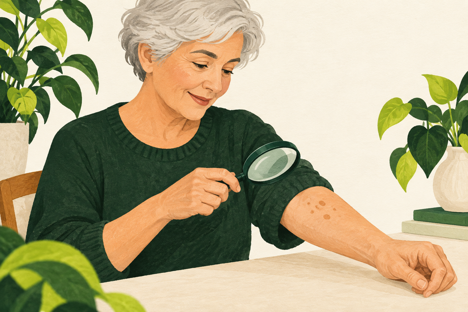
AI-generated conceptual illustration; not a diagnostic image or depiction of a real patient.
A raised, rough spot that looks pasted onto the skin may be a seborrheic keratosis. These common growths are benign, but that reassurance applies to a correctly identified lesion. A photograph, a search result, or the fact that a spot appeared with age cannot reliably distinguish every harmless growth from skin cancer.
What a seborrheic keratosis can look like
The surface may look waxy, warty, or crumbly. Color varies considerably, from pale shades to brown or nearly black. Some begin as small rough bumps and gradually become thicker. They commonly occur on the trunk, face, scalp, or neck and may be solitary or numerous. Many cause no symptoms; others itch or catch on clothing. The American Academy of Dermatology describes this range of appearances.
Those features are useful background, not a home diagnostic checklist. A dark growth is not automatically dangerous, and a familiar-looking one is not automatically safe. What matters is whether its identity is clear and whether it is changing.
Why they appear
Seborrheic keratoses become more common in middle and later life, and a tendency to develop them can run in families. They occur across skin tones. The cause is not fully understood. Sun exposure may contribute, but growths also develop on covered skin. They are not contagious, so new spots appearing elsewhere do not mean you have spread an infection by touching them. AAD’s explanation of causes acknowledges this uncertainty.
The British Association of Dermatologists also distinguishes these growths from viral warts and notes that confirmed seborrheic keratoses do not become skin cancer. Their name should not be taken to mean an oil-gland problem or an infection requiring an antiseptic.
Changes worth checking
Arrange a medical assessment if a growth enlarges quickly, changes color, bleeds, becomes persistently itchy or sore, or looks different from your other spots. A sudden crop of many new growths also deserves discussion. This does not establish a cancer diagnosis; it is a reason to avoid assuming the cause. A flat, rough, scaly patch might instead be an actinic keratosis, which needs different advice. These are among the AAD’s reasons to see a dermatologist.
For the appointment, note when you first noticed the lesion and what has changed. A dated photograph can help explain the timeline, but should not delay assessment. Mention repeated rubbing from a bra strap, collar, jewelry, or shaving because irritation may influence the treatment discussion.
Observation or removal?
If a clinician is confident that a growth is a seborrheic keratosis and it causes no trouble, leaving it alone is reasonable. If the diagnosis is uncertain, a small tissue sample or removal for microscopic examination may be needed. Destructive cosmetic treatment should not come before that decision.
Removal options include freezing with liquid nitrogen, scraping the growth away, or using an electrical procedure, sometimes in combination. The choice depends on the lesion and clinical circumstances. Treatment can leave a lighter patch, and color changes may sometimes persist. Ask about the expected appearance, healing, and aftercare before agreeing to removal. AAD’s treatment guidance explains these options and their limitations.
Removing one lesion does not prevent new ones from appearing elsewhere, and a treated lesion may occasionally return. Cosmetic improvement and relief from friction are useful outcomes; neither is a guarantee that your skin will remain free of future growths.
Do not remove an uncertain spot at home
Do not cut, burn, scrape, or apply wart-removal products to a presumed seborrheic keratosis yourself. Besides infection and injury, self-treatment can interfere with getting the right diagnosis. Reduce avoidable rubbing while arranging care rather than trying to erase the spot first.
Evidence note
Sources were checked on October 13, 2026. The AAD treatment page was updated in January 2023; the BAD leaflet is dated October 2022, with a listed review due in 2025. These are professional patient-education sources, not a head-to-head trial proving one removal method best. No remote description can confirm the identity of an individual lesion.
Editorial disclosure: AI assisted with research, writing and the conceptual illustration. This is general health information, not personalized medical advice.